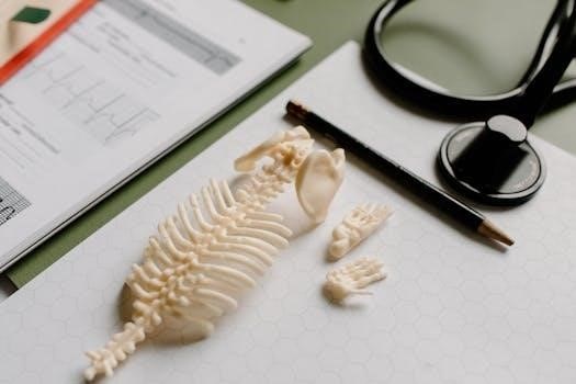
Welcome to your comprehensive skeletal system study guide! This resource is designed to help you navigate the complexities of the human skeleton. From its functions to its intricate bone structures, we’ll explore the axial and appendicular divisions, ensuring a solid understanding.
Functions of the Skeletal System
The skeletal system is far more than just a framework; it’s a dynamic and essential component of the human body, performing a multitude of vital functions. Primarily, it provides support, acting as a scaffold that upholds our body’s structure and maintains its shape.
Closely linked to support is protection. Bones like the skull and rib cage safeguard delicate organs such as the brain, heart, and lungs from injury. This protective role is crucial for survival.

The skeletal system facilitates movement. Bones serve as levers, and joints act as fulcrums, allowing muscles to generate motion. This interaction enables a wide range of physical activities.
Beyond structural roles, the skeletal system is involved in storage. Bones store essential minerals like calcium and phosphate, releasing them into the bloodstream when needed. This helps maintain mineral balance in the body.
Another vital function is blood cell production, or hematopoiesis. Red bone marrow, found within certain bones, produces red blood cells, white blood cells, and platelets, all critical for oxygen transport, immune response, and blood clotting.
Axial Skeleton⁚ Skull, Vertebral Column, and Bony Thorax
The axial skeleton forms the central, longitudinal axis of the body, providing support, protection, and attachment points for the appendicular skeleton. It comprises three major components⁚ the skull, vertebral column, and bony thorax.
The skull, the most complex part of the axial skeleton, is composed of cranial and facial bones. The cranium protects the brain, while the facial bones form the face, house sensory organs, and provide attachment points for facial muscles.
The vertebral column, or spine, extends from the skull to the pelvis. It supports the head and trunk, protects the spinal cord, and allows for movement and flexibility. It consists of cervical, thoracic, lumbar, sacral, and coccygeal vertebrae.
The bony thorax, or rib cage, protects the organs of the thoracic cavity, including the heart and lungs. It is formed by the sternum, ribs, and thoracic vertebrae. The rib cage expands and contracts during breathing, aiding in respiration.
These three components work together to provide a stable, protective framework for the body’s central axis, enabling essential functions and movements. Understanding their individual structures and relationships is crucial for comprehending the overall function of the skeletal system.
Skull⁚ Cranium and Facial Bones
The skull, the most complex bony structure in the body, is divided into two main sections⁚ the cranium and the facial bones. The cranium, composed of eight bones, encloses and protects the delicate brain tissue. These bones are joined together by sutures, which are immovable joints that provide stability to the skull. The cranial bones include the frontal, parietal (paired), temporal (paired), occipital, sphenoid, and ethmoid bones.
The facial bones, fourteen in total, form the framework of the face, providing structure for the eyes, nose, and mouth. These bones contribute to our unique facial features and provide attachment points for facial muscles. The facial bones include the nasal (paired), zygomatic (paired), maxillary (paired), mandible, lacrimal (paired), palatine (paired), inferior nasal conchae (paired), and vomer bones.

Together, the cranium and facial bones form a protective and supportive structure for the head, allowing for sensory perception, facial expression, and the initial stages of digestion through chewing. Understanding the individual bones and their functions is essential for comprehending the overall structure and function of the skull.
Vertebral Column⁚ Cervical, Thoracic, and Lumbar Vertebrae
The vertebral column, also known as the spine, is a flexible, S-shaped structure that supports the head, neck, and trunk. It extends from the skull to the pelvis and is composed of 33 individual vertebrae, though some fuse during development. These vertebrae are divided into five regions⁚ cervical, thoracic, lumbar, sacral, and coccygeal.
The cervical vertebrae, located in the neck, consist of seven vertebrae (C1-C7). They are the smallest and most mobile vertebrae, allowing for a wide range of head movements. The first two cervical vertebrae, the atlas (C1) and axis (C2), are specialized for head rotation and nodding.
The thoracic vertebrae, located in the upper back, consist of twelve vertebrae (T1-T12). They articulate with the ribs, forming the rib cage. These vertebrae are less mobile than the cervical vertebrae due to their articulation with the ribs.
The lumbar vertebrae, located in the lower back, consist of five vertebrae (L1-L5). They are the largest and strongest vertebrae, bearing the majority of the body’s weight. These vertebrae have thick, block-like bodies and are adapted for weight-bearing and flexibility. Understanding the regional differences in vertebral structure is essential for comprehending the function and biomechanics of the spine.

Appendicular Skeleton⁚ Limbs and Girdles
The appendicular skeleton comprises the bones of the limbs and the girdles that attach them to the axial skeleton. It is responsible for locomotion and manipulation of the environment. This system includes the pectoral girdle, upper limbs, pelvic girdle, and lower limbs, all working together to enable movement and interaction with our surroundings.
The pectoral girdle, consisting of the clavicle and scapula, connects the upper limbs to the axial skeleton, providing flexibility and range of motion. The upper limbs include the humerus, radius, ulna, carpals, metacarpals, and phalanges, forming the arm, forearm, and hand. These bones allow for precise movements and grasping abilities.
The pelvic girdle, formed by the two hip bones (ossa coxae), connects the lower limbs to the axial skeleton, providing stability and weight-bearing support. The lower limbs consist of the femur, tibia, fibula, tarsals, metatarsals, and phalanges, forming the thigh, leg, and foot. These bones are designed for weight-bearing, balance, and propulsion during locomotion.
Together, the appendicular skeleton’s components enable a wide array of movements and provide the framework for interacting with the world around us. Understanding the structure and function of these bones is crucial for comprehending human movement and biomechanics.
Pectoral Girdle⁚ Clavicle and Scapula
The pectoral girdle, also known as the shoulder girdle, is responsible for connecting each upper limb to the axial skeleton. It consists of two bones⁚ the clavicle and the scapula. These bones work in tandem to provide a wide range of motion and flexibility to the shoulder joint, enabling various arm movements.
The clavicle, or collarbone, is a slender, S-shaped bone that acts as a strut between the scapula and the sternum. It helps to transmit forces from the upper limb to the axial skeleton, preventing shoulder dislocation. The clavicle also provides attachment points for muscles and ligaments, contributing to shoulder stability.
The scapula, or shoulder blade, is a flat, triangular bone located on the posterior aspect of the thorax. It articulates with the humerus at the glenohumeral joint (shoulder joint) and with the clavicle at the acromioclavicular joint. The scapula provides attachment points for numerous muscles that control shoulder movement and stability.
Together, the clavicle and scapula form the pectoral girdle, allowing for a wide range of upper limb movements, including abduction, adduction, flexion, extension, rotation, and circumduction. Their unique structure and articulation contribute to the shoulder’s mobility and functionality.
Upper Limb⁚ Humerus, Radius, Ulna, Carpals, Metacarpals, Phalanges
The upper limb, responsible for a vast array of movements and manipulations, is comprised of 30 distinct bones. These bones form the skeletal framework of the arm, forearm, and hand, each playing a crucial role in the limb’s overall function. The upper limb begins with the humerus, a long bone that extends from the shoulder to the elbow. At the elbow, the humerus articulates with the radius and ulna, the two bones of the forearm.
The radius is located on the lateral side of the forearm, while the ulna is positioned medially. These bones work together to allow for pronation and supination of the forearm. Distally, the radius and ulna connect to the carpal bones, which form the wrist. The carpals are a group of eight small bones arranged in two rows.

Beyond the wrist lie the metacarpals, five bones that form the palm of the hand. Each metacarpal articulates with a proximal phalanx, one of the bones of the fingers. Each finger has three phalanges – proximal, middle, and distal – except for the thumb, which has only two. This intricate arrangement of bones allows for the fine motor skills and grasping abilities that characterize the human hand.
Pelvic Girdle⁚ Hip Bones
The pelvic girdle, a crucial component of the skeletal system, is primarily formed by two coxal bones, also known as ossa coxae or more commonly, hip bones. These bones provide a strong and stable base for the attachment of the lower limbs to the axial skeleton. Each hip bone is a fusion of three distinct bones⁚ the ilium, the ischium, and the pubis.
During childhood, these bones are separate, but they gradually fuse together to form a single, solid structure by adulthood. The ilium is the largest of the three bones and forms the superior part of the hip bone. The ischium forms the posteroinferior part of the hip bone and is characterized by the ischial tuberosity, which bears the weight of the body when sitting.
The pubis forms the anterior part of the hip bone. The two pubic bones articulate at the pubic symphysis, a cartilaginous joint that connects the left and right sides of the pelvic girdle. Together, the hip bones, along with the sacrum and coccyx of the vertebral column, form the bony pelvis, a basin-shaped structure that supports the torso, protects the pelvic organs, and provides attachment points for numerous muscles.
Lower Limb⁚ Femur, Tibia, Fibula, Tarsals, Metatarsals, Phalanges
The lower limb is designed for weight-bearing and locomotion, comprising the thigh, leg, and foot. The femur, or thigh bone, is the longest and strongest bone in the human body. It articulates with the acetabulum of the pelvis, forming the hip joint, and with the tibia at the knee joint. Distally, the lower leg is composed of two bones⁚ the tibia and fibula.
The tibia, or shinbone, is the larger and more medial bone, bearing most of the weight. The fibula, located laterally, is thinner and primarily serves for muscle attachment and stabilization of the ankle. These bones are connected by an interosseous membrane. The foot consists of the tarsals, metatarsals, and phalanges.
The tarsals, comprising seven bones, form the ankle and posterior part of the foot, including the calcaneus (heel bone). Five metatarsals form the arch of the foot, connecting the tarsals to the phalanges. Each toe consists of phalanges, with the hallux (big toe) having two and the others having three. These bones enable balance, propulsion, and shock absorption during movement.
Joints or Articulations⁚ Fibrous, Cartilaginous, and Synovial
Joints, also known as articulations, are crucial for connecting bones and facilitating movement. They provide stability and mobility, allowing the skeleton to function effectively. Joints are classified structurally based on the material that binds the bones together⁚ fibrous, cartilaginous, and synovial.
Fibrous joints are characterized by bones united by fibrous tissue, resulting in limited or no movement. Examples include sutures in the skull. Cartilaginous joints feature bones connected by cartilage, allowing for slight movement. These include synchondroses (hyaline cartilage, like the epiphyseal plates) and symphyses (fibrocartilage, like the intervertebral discs).
Synovial joints are the most common type, distinguished by a joint cavity containing synovial fluid, which reduces friction and provides nourishment. These joints, predominantly found in the limbs, permit a wide range of motion. Synovial joints include plane, hinge, pivot, condyloid, saddle, and ball-and-socket joints, each offering unique movement capabilities based on the articulating bone surfaces’ shapes, crucial for diverse activities.