Understanding Tennis Elbow (Lateral Epicondylopathy)
Lateral elbow tendinopathy (LET), commonly known as tennis elbow, affects adults aged 40-60, with an annual incidence of 1-3%. Microtrauma and repetitive strain
cause lesions at the extensor carpi radialis brevis (ECRB) origin, leading to degenerative tendon changes.
Definition and Commonality
Lateral epicondylopathy (LET), frequently called tennis elbow, represents the most prevalent cause of elbow pain in adult populations, specifically those between 40 and 60 years of age. Its annual incidence ranges from 1 to 3 percent, highlighting its significant impact on individuals within this demographic. This condition isn’t exclusive to tennis players; any activity involving repetitive wrist extension and forearm rotation can contribute to its development.
At its core, LET is a degenerative condition affecting the tendons on the outer elbow. The primary lesion typically occurs at the origin of the extensor carpi radialis brevis (ECRB) muscle, though the extensor carpi radialis longus (ECRL) and extensor digitorum muscles can also be involved. The pathology arises from microtrauma and repetitive injury, leading to gradual tendon degeneration rather than acute inflammation. Prolonged contraction and tension within the forearm extensor muscles exacerbate these degenerative changes over time.
The repetitive nature of actions like gripping, lifting, and even typing can contribute to the development of LET, making it a common ailment across various professions and lifestyles. Understanding its definition and prevalence is crucial for effective prevention and management strategies.
Causes and Risk Factors (Repetitive Strain)
The primary cause of lateral elbow tendinopathy (LET) is repetitive strain on the forearm extensor muscles, particularly the extensor carpi radialis brevis (ECRB). This strain stems from activities involving repeated wrist extension, supination, and forceful gripping. While termed “tennis elbow,” the condition frequently arises from non-athletic pursuits like plumbing, carpentry, or even prolonged computer use.
Several risk factors heighten susceptibility to LET. These include occupations demanding repetitive arm movements, improper technique during sports (especially racquet sports), and inadequate conditioning of forearm muscles. Increasing age also plays a role, as tendons lose elasticity over time, making them more vulnerable to injury. Individuals with poor wrist mechanics or those who suddenly increase activity levels are also at increased risk.
The underlying mechanism involves microtrauma to the tendon’s origin, leading to small tears that accumulate over time. Long-term contraction and tension within the forearm extensors contribute to degenerative changes, ultimately resulting in pain and functional limitations. Addressing these risk factors is vital for both prevention and rehabilitation.
Anatomy of Affected Tendons (ECRB, ECRL, Extensor Digitorum)

Lateral elbow tendinopathy (LET) primarily affects the tendons responsible for wrist extension. The extensor carpi radialis brevis (ECRB) is most commonly implicated, stabilizing the wrist during elbow extension and being particularly vulnerable due to its insertion point. However, the extensor carpi radialis longus (ECRL) and extensor digitorum can also contribute to the pathology.

These tendons originate from a common extensor tendon at the lateral epicondyle of the humerus. The ECRB’s insertion is on the second metacarpal bone, while the ECRL inserts on the third metacarpal. The extensor digitorum extends not only the wrist but also the fingers. Understanding their anatomical relationships is crucial for targeted treatment.
Repetitive stress leads to degeneration at the tendons’ origin, causing pain radiating down the forearm. The ECRB’s susceptibility stems from its role in forceful wrist extension and supination, common movements in activities triggering LET. Proper diagnosis requires recognizing the specific tendons involved and the extent of the damage.
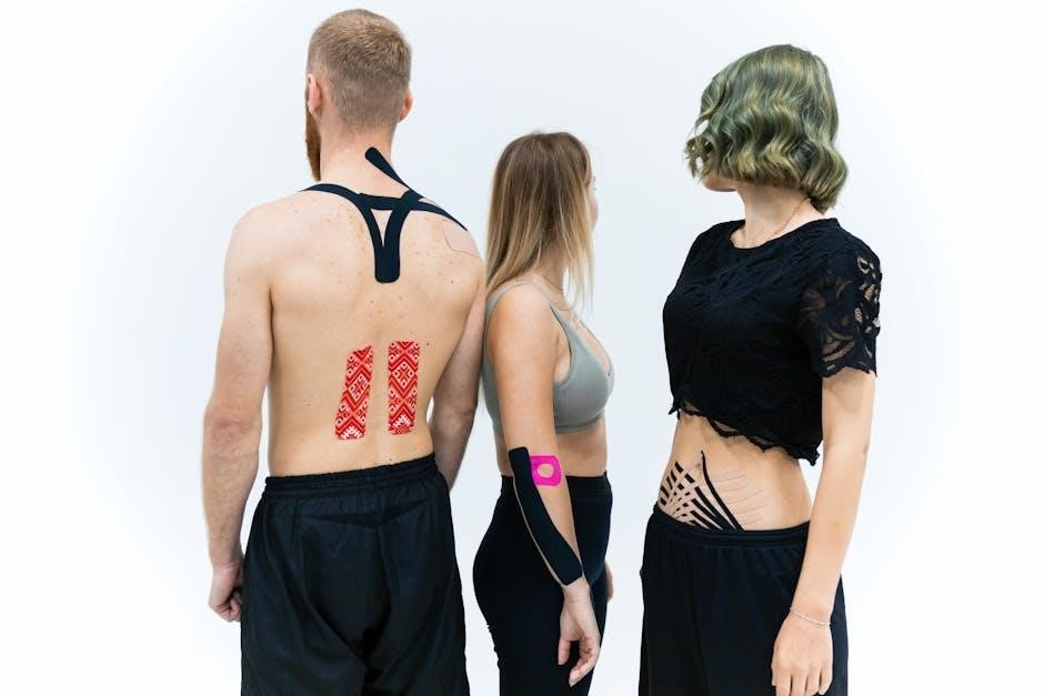
Kinesio Taping (KT) is a rehabilitative taping technique based on the body’s natural healing processes. It aims to reduce pain, support muscles, and correct fascial dysfunction, potentially aiding LET recovery.
What is Kinesio Taping?
Kinesio Taping is a therapeutic method employing an elastic adhesive tape, differing significantly from traditional athletic taping. Developed by Kenzo Kase, it’s designed to mimic the elasticity of human skin, allowing for a full range of motion while providing support. Unlike restrictive taping which limits movement, Kinesio Tape facilitates it, enabling continued activity during rehabilitation.
The tape itself is thin, lightweight, and breathable, often made of a cotton-elastane blend. It doesn’t provide rigid immobilization; instead, it gently lifts the skin, creating space between the skin and underlying tissues. This decompression is believed to influence the body’s natural healing mechanisms.

Kinesio Taping isn’t a cure, but rather a tool to assist the body’s own recovery processes. It’s frequently used in conjunction with other therapies, such as exercise and manual therapy. The application technique is crucial, and a trained professional should apply the tape to achieve optimal results. Different application methods target various physiological effects, making it a versatile therapeutic option.
How Kinesio Tape Works (Pain Reduction, Muscle Support, Fascial Correction)
Kinesio Tape exerts its therapeutic effects through several interconnected mechanisms. Primarily, it’s believed to reduce pain by influencing the nervous system. Lifting the skin with the tape creates space, potentially decreasing pressure on pain receptors and modulating pain signals sent to the brain.
Furthermore, the tape provides dynamic support to muscles without restricting their full range of motion. This support can enhance muscle function and reduce strain on injured tissues. For conditions like tennis elbow, it assists the extensor muscles, allowing them to function more efficiently during activity.
A key aspect is fascial correction. The tape can influence the fascia, the connective tissue network surrounding muscles. By gently lifting and repositioning the fascia, it can improve tissue glide, reduce adhesions, and restore optimal biomechanics. In the context of tennis elbow, this helps correct imbalances in the forearm muscles and alleviate tension at the affected tendon origin.

Kinesio Taping Techniques for Tennis Elbow
Kinesio taping for lateral epicondylitis utilizes two Y-shaped strips. One strip supports extensor muscles, while the second corrects fascia, applied with the elbow extended and wrist flexed.
Materials Needed (Kinesio Tape, Scissors)
Successfully applying Kinesio Tape for tennis elbow requires minimal, readily available materials. The cornerstone of the process is, naturally, a roll of high-quality Kinesio Tape itself. Ensure the tape is appropriate for sensitive skin, especially if the patient has any allergies or sensitivities to adhesives. The width of the tape commonly used for this application is 2 inches (5cm), providing sufficient coverage for the targeted muscle groups and adequate surface area for effective adhesion.
Alongside the tape, a pair of sharp, rounded-tip scissors is essential. These are needed to precisely cut the tape into the required shapes – specifically, the Y-strips utilized in the application technique. Rounded tips are crucial for safety, preventing accidental skin punctures during the cutting process. Avoid using standard pointed scissors, as they pose a higher risk of injury.
While not strictly required, additional materials can enhance the application. These include skin pre-spray to remove oils and lotions for better adhesion, and athletic tape to secure the ends of the Kinesio Tape, particularly for patients with high activity levels. However, the core necessities remain the Kinesio Tape and the scissors.
Patient Positioning (Elbow Extended, Wrist Flexed & Ulnar Deviated)
Proper patient positioning is paramount for effective Kinesio Taping application in treating tennis elbow. Begin by having the patient sit comfortably with their affected arm supported, allowing for complete relaxation of the forearm muscles. The elbow should be fully extended – straight – but not hyperextended, maintaining a natural anatomical alignment. This position ensures the extensor muscles are at their optimal length for tape application.
Next, instruct the patient to gently flex their wrist, bending it downwards as if signaling “stop”. Simultaneously, they should perform ulnar deviation, moving their hand towards the little finger side. This combined movement places the wrist in a position that effectively stretches and isolates the extensor muscles responsible for tennis elbow pain – specifically, the extensor carpi radialis brevis (ECRB).
Maintaining this position throughout the taping process is crucial. It allows for accurate tape placement along the muscle fibers and ensures the desired tension is applied correctly. Consistent positioning maximizes the tape’s ability to provide support, reduce pain, and facilitate proper muscle function.
Application of the First Y-Strip (Along Extensor Muscles ‒ 30% Tension)
Begin with a Y-shaped Kinesio tape strip. The tail ends should be approximately half the length of the stem. Anchor the base (stem) of the ‘Y’ on the patient’s wrist, slightly towards the thumb side, with no tension – this is the anchor point. Ensure the patient maintains the previously established elbow extension, wrist flexion, and ulnar deviation.
Apply a gentle, consistent 30% tension to the tape tails as you follow the path of the forearm extensor muscles. This means stretching the tape to approximately 30% of its maximum elongation capacity. Guide the tails proximally (towards the elbow), adhering the tape along the bellies of the extensor carpi radialis brevis (ECRB) and other relevant extensor muscles.
Avoid excessive tension, as this can restrict movement and potentially exacerbate discomfort. The goal is to provide support without compromising function. Once the tails are applied, gently rub the tape to activate the heat-sensitive adhesive, ensuring secure adhesion to the skin. Finish by applying no tension to the very ends of the tape.
Application of the Second Strip (Vertical to First ౼ Fascial Correction)
Utilize a second Kinesio tape strip, typically an ‘I’ strip, with a width similar to the first. Begin by anchoring this strip with zero tension directly over the affected area on the proximal forearm – perpendicular to the previously applied Y-strip. This placement aims to address fascial restrictions contributing to the condition.
Apply a gentle tension – around 10-20% – as you guide the tape vertically, crossing over the first Y-strip. The purpose of this strip isn’t muscle inhibition, but rather fascial correction. It helps to lift the fascia, improving space and reducing compression around the affected tendons and muscles.
Ensure the tape follows a straight path, avoiding any twisting or bunching. Gently rub the tape after application to activate the adhesive and promote secure attachment to the skin. This second strip works synergistically with the first, providing a more comprehensive approach to addressing the biomechanical imbalances associated with lateral epicondylopathy.

Evidence-Based Research on Kinesio Taping for Tennis Elbow
Research indicates that Kinesio taping (KT) can effectively reduce pain and enhance function in patients experiencing lateral elbow tendinopathy (LET). Studies show clinically significant improvements in VAS and PRTEE scores.
Meta-Analysis Findings (Pain Reduction & Function Improvement)
Meta-analyses examining the effects of Kinesio taping (KT) on lateral elbow tendinopathy (LET) consistently demonstrate positive outcomes regarding pain reduction and functional improvements. These comprehensive reviews synthesize data from multiple studies, providing a stronger level of evidence than individual trials alone. Findings suggest that KT is a viable adjunct therapy for managing LET symptoms.
Specifically, the analyses reveal statistically significant decreases in pain intensity, as measured by the Visual Analog Scale (VAS), following KT application. Patients report a noticeable lessening of discomfort during both activity and rest. Furthermore, improvements are observed in functional capacity, assessed using tools like the Patient-Rated Tennis Elbow Examination (PRTEE). This indicates that KT helps individuals return to daily activities with greater ease and reduced limitations.
The mechanisms underlying these benefits are thought to involve pain modulation, enhanced muscle support, and correction of fascial imbalances. While the exact contribution of each mechanism is still under investigation, the collective evidence supports the use of KT as a conservative treatment option for LET. However, it’s crucial to acknowledge the limitations of current research and the need for further high-quality studies.
VAS and PRTEE Score Improvements
Studies consistently report clinically significant improvements in both Visual Analog Scale (VAS) and Patient-Rated Tennis Elbow Examination (PRTEE) scores following Kinesio Taping (KT) treatment for lateral elbow tendinopathy (LET). The VAS, a common pain measurement tool, demonstrates a reduction in pain intensity among patients receiving KT, indicating a tangible decrease in perceived discomfort.
The PRTEE, a patient-reported outcome measure, assesses functional limitations associated with LET. Research shows that KT application leads to notable enhancements in PRTEE scores, signifying improved ability to perform daily activities and participate in recreational pursuits. These improvements suggest KT positively impacts a patient’s overall quality of life.
Specifically, observed changes in VAS scores often exceed minimal clinically important differences, meaning the pain reduction is noticeable and meaningful to patients. Similarly, PRTEE score improvements reflect a substantial lessening of functional impairments. These quantifiable outcomes provide objective evidence supporting the efficacy of KT as a therapeutic intervention for LET, although further rigorous research is still warranted to confirm these findings and optimize application techniques.

Limitations of Current Research & Need for Further Studies
Despite promising findings, current research on Kinesio Taping (KT) for lateral elbow tendinopathy (LET) exhibits several limitations. Many studies suffer from small sample sizes, hindering the generalizability of results. Methodological inconsistencies, including variations in KT application techniques and assessment protocols, also contribute to difficulty in comparing findings across studies.
A lack of robust control groups, such as sham taping or standardized exercise programs, often limits the ability to definitively attribute observed improvements solely to KT. Furthermore, the long-term effects of KT remain largely unknown, with few studies investigating sustained benefits beyond the immediate post-treatment period.
Therefore, future high-quality studies employing rigorous protocols are crucial. These should include larger, randomized controlled trials with well-defined inclusion criteria, standardized KT application procedures, and long-term follow-up assessments. Investigating the optimal taping parameters – tension, strip width, and application duration – is also essential to maximize KT’s therapeutic potential and establish evidence-based guidelines for clinical practice.